Written by Philippos Rahal, Οptician – Optrometrist
Dry eye condition is quite often in our days. Actually, it has a lot to do with the modern way of life because It’s a multi factor condition.
So, we are going to make a list of the tests, assessing dry eye. According to TFOS DEWS II there are two basic categories of the tests, invasive and non-invasive.
Invasive Tests
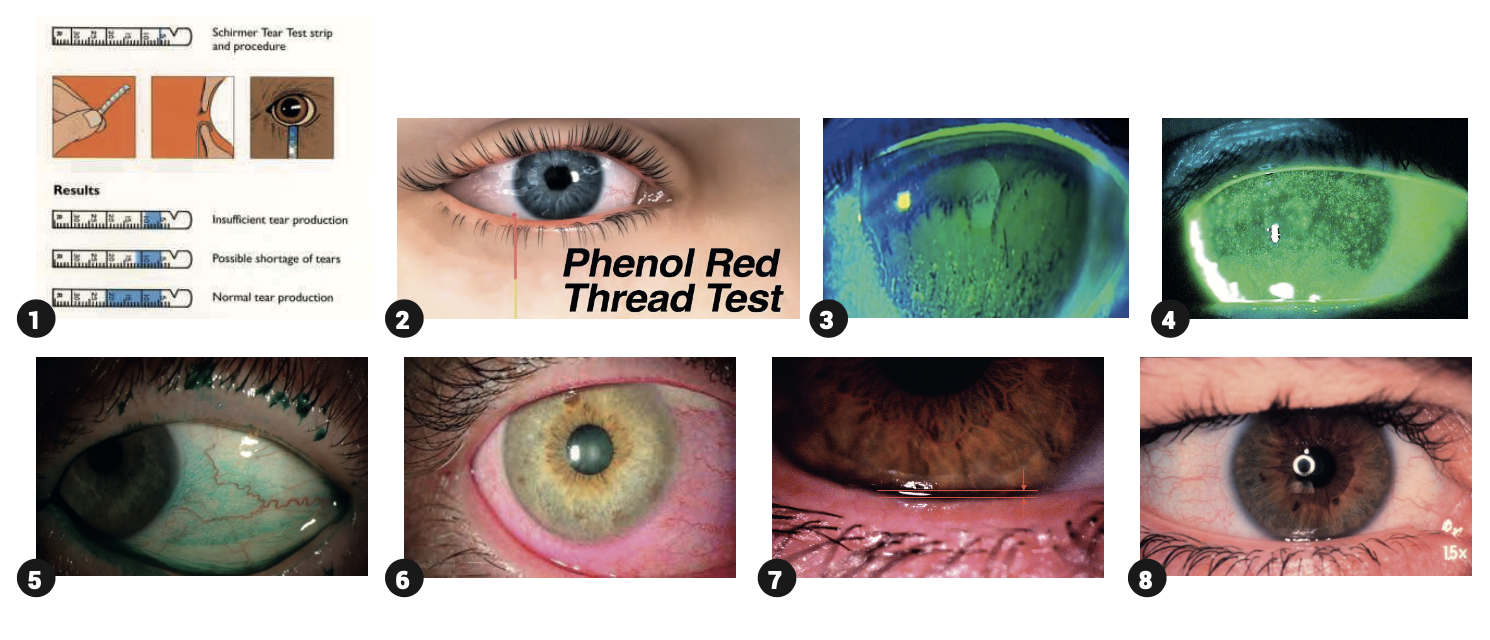
Schirmer & Phenol Red test: Phenol Red test is less irritating for the patient and much shorter in its execution.
Break Up Time (BUT): Use of fluorescein.
Ocular Surface assessing: Use of fluorescein.
Lissamine Green & Rose Bengal: Lissamine – Green is less irritating for the ocular surface.
Corneal Sensitivity: Assess is empirical in this case.
It’s important to dictate that with all these invasive tests, that are been executed on the slit lamp, there is an extra quite annoying factor and that is its luminance.
Non-Invasive Tests
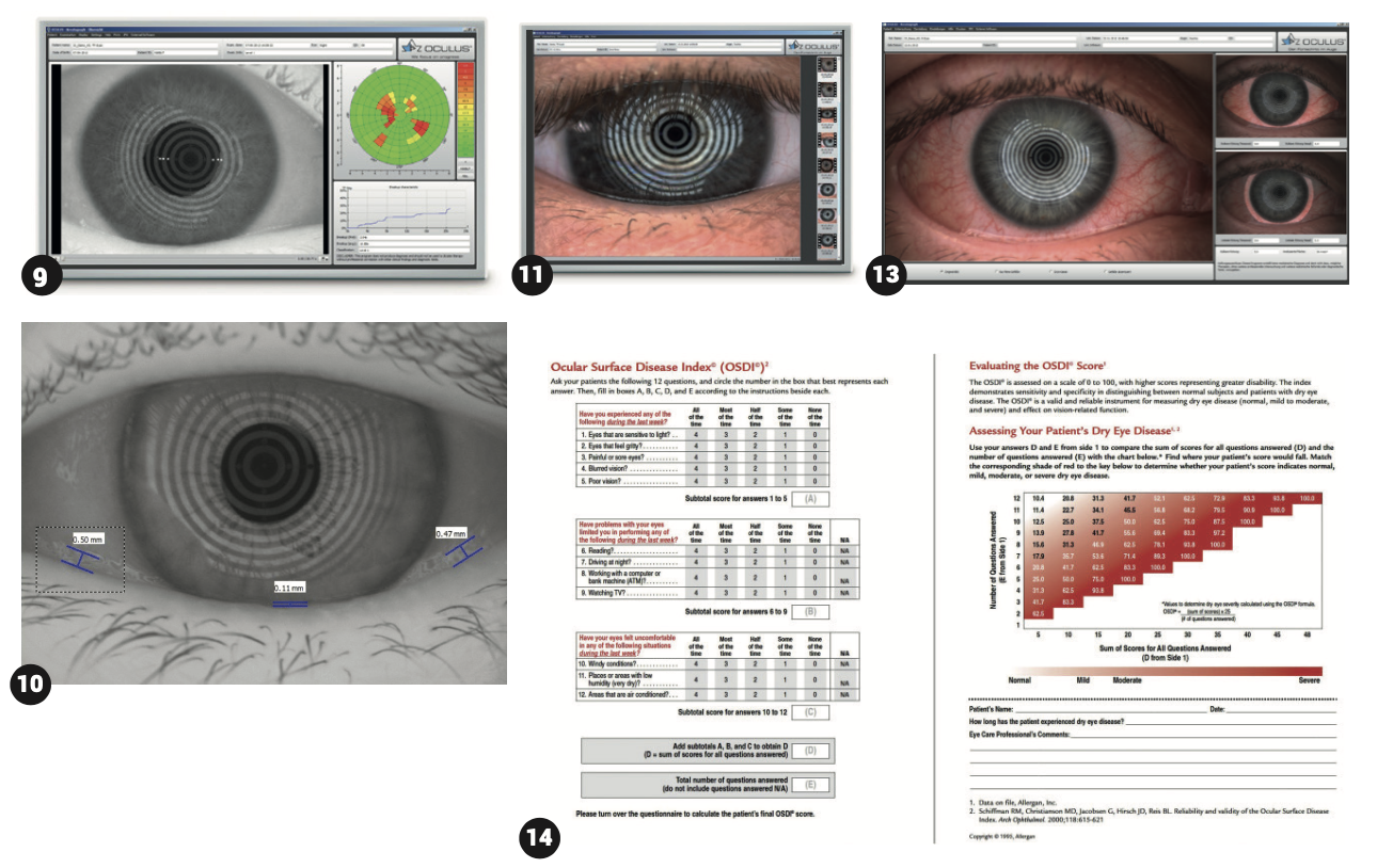
Measurement of the Tear Meniscus: Use of the slit lamp.
Lipid layer assessment: Use of the slit lamp.
Tear Film quality: Using the slit lamp we can assess the tear film’s viscosity, whether is low or high.
Redness of the conjunctiva: examination of the conjunctival vessels while determinating whether they are conjunctival or/and scleral using the grading scale at the same time.
Non-Invasive Break Up Time (NBUT): using Dry Eye Analyzer (DEA) devices
Tear Meniscus Assessment using DEA: measurement of the height of tear meniscus.
Assessment of the Lipid Layer using DEA: examination of the lipid layer status, whether it exists and in which percentage.
Assessment of aqueous layer Debris using DEA: while focusing on the aqueous layer of the tear film.
Assessment of the Conjunctival Redness using DEA: the whole ocular surface is been photographed by the device. At the same time, it can focus and assess the temporal or/nasal area.
Dry Eye Questionnaires: they can quantify the dry eye status of the patient and its impact to his daily life.
Assessment of the Meibomian glands on upper and lower eyelids: It’s one of the most critical tests in order to determine the type of any specific dry eye condition. In case of high rate of meibomian gland disfunction (MGD) the direction of the disease is mainly evaporative. On the other hand, it could be due to less aqueous production through the lacrimal gland, for example in Sjogren syndrome. In many cases the dry eye condition is mixed.
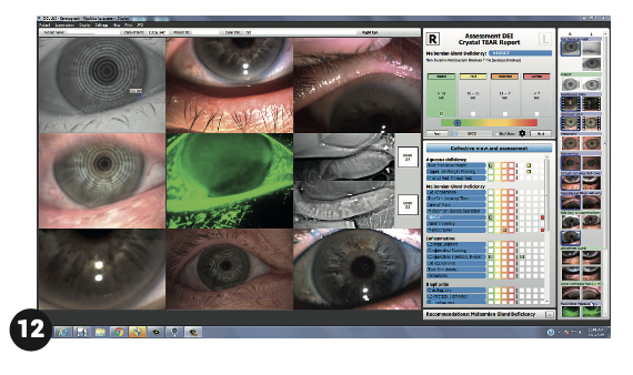
For the MGD assessment it is necessary to perform eversion of the eyelids both upper and lower ones. We can do that while using the slit lamp or a DEA. Through these devices all tests can be saved, assessed, compared to each other and of course categorized according to the TFOS DEWS II grading system.
Latest information regarding to Dry Eye condition
- MMP-9 (Matrix Metalloproteinase-9) specific devices we can assess the matrix metalloproteinase-9 concentration in the tear film.
- Osmolarity of the tear film: using specific devices we can assess again the tear film osmolarity.
A normal value ranges from 302mOsm/L (+/-8), a moderate from 315mOsm/L (+/-10) and a severe one from 322mOsm/L (+/-22). All these values show the grade of DE, the greater the value is, the higher the ocular surface disease is. (speaking of DE). In general, a value higher from 308mOsm/L is considered a threshold to DE condition.
• Optical Coherence Tomography (OCT): using the OCT (anterior chamber) we can acquire the epithelium map of the entire cornea. Epithelium changes are the first to appear when DE condition starts and then evolves.
• FVA (Functional Visual Acuity): It is about the time in between two blinks until the quality of vision or/and the visual acuity deteriorates. Usually, it has to do with practical issues of the everyday life, of the patient.
TFI (Tear Function Index): after having assessed the tear production (Schirmer Test) then we observe it’s drainage, in order to extract a conclusion of the DE severity.
Lid Wiper Epitheliopathy (LWE): using Lissamine Green we can achieve eye lids edge staining. With this method we can assess if the tear film is been outspread in the whole ocular surface.
Dry eye is often enough in order for us to consider it in every patient we exam.
It is not necessary though to perform every test we know. Combining the patient’s history and 2 or 3 tests, we have a really wide picture of what we are dealing with in each case.


